+91 9393 108 108
- Our Doctors
- Our Specialities
Centres of Excellence
-
 Centre for Blood Diseases, BMT & Cancer Immunotherapy
 -
 Centre for Bone, Joint & Spine
 -
 Centre for Critical Care Medicine and ECMO Services
 -
 Centre for Gastrosciences
 -
 Centre for Heart & Vascular Care
 -
 Centre for Nephro-Urosciences
 -
 Centre for Neurosciences
 -
 Centre for Obstetrics and Gynaecology
 -
 Centre for Organ Transplantation
Super Speciality
-
 Advanced Diagnostic and Interventional Radiology
 -
 Anesthesiology & Pain Management
 -
 Clinical Nutrition and Dietetics
 -
 Dental and Maxillofacial Surgery
 -
 Dermatology
 -
 Emergency and Trauma
 -
 Endocrinology and Metabolic Disease
 -
 ENT and Head & Neck Surgery
 -
 Family Medicine
 -
 General and Laparoscopic Surgery
 -
 General Medicine
 -
 Laboratory Medicine
-
- Key Procedures
- Our Hospitals
- International Patient
- Contact us
-
Quick Links


Blogs
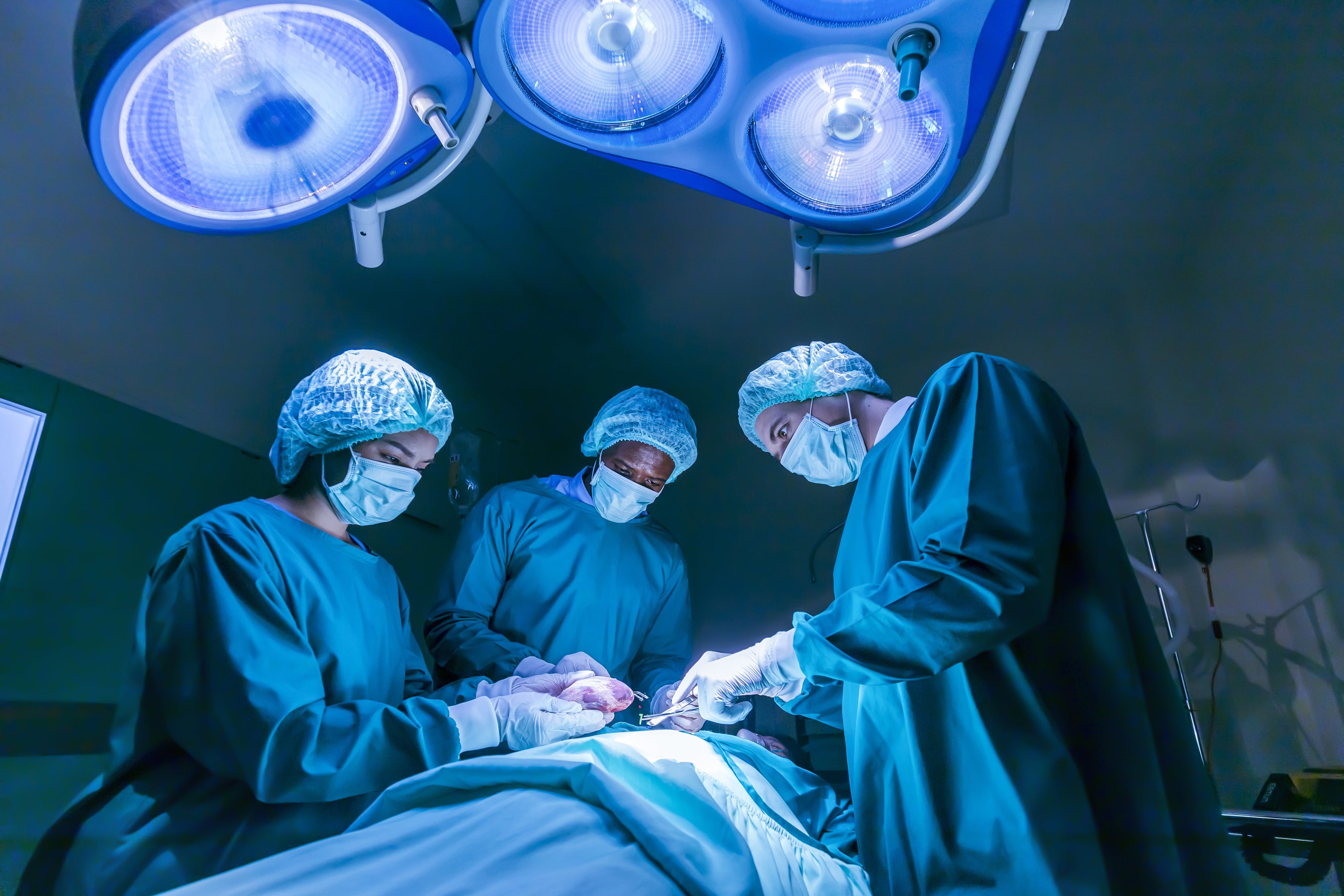
Awake Craniotomy
An awake craniotomy is an operation performed in the same manner as a conventional craniotomy( brain surgery) but with the patient awake during the critical part of the procedure.
This is a preferred technique for operations to remove tumours close to, or involving functionally important (eloquent) regions of the brain. Performing the operation in this way allows us to test regions of the brain before they are incised or removed and also to test the patient’s function continuously throughout the operation. The overall aim is to minimise the risks of the operation.
The benefits are considered to be of increased extent of lesion removal, with growing evidence of improved survival benefit, whilst minimizing damage to eloquent cortex and resulting postoperative neurological dysfunction. Other advantages include a shorter hospitalization time, hence reduced cost of care, and a decreased incidence of post-anesthesia complications such as nausea and vomiting.
Not all patients are fit for awake craniotomy. A team of neurologists/ neurosurgeons/ neuropsychologists/ anesthesiologists and neurophysiologists together select the right candidate. Thorough pre-operative assessment of airway and other premorbid conditions like sleep apnea, mental impairment, personality disorder, pre-existing paralysis, brain swelling and profound dysphasia is essential.
The team members,especially the anesthetistist, must gain the patient’s confidence, as the patient will depend on them during the procedure. Prior to surgery, the patient must be informed about realistic description of the operating room, expected discomforts and level of co-operation expected, potential risks, safety measures and stages of the procedure. The anesthesiologist must explain in detail the potential presence of sounds (monitor alarms, cranial drilling, elektroknife, ultrasonic surgical aspirator) or discomforts (unchangeable position, aphasia during cortical mapping) from the patient. The patient must understand that these discomforts are essential for the success of the procedure. A visit to the operating room before surgery in order to familiarize the patient with the sounds and equipment in the rooms is a good idea. The patient should be explained the tasks that will be performed for speech and motor testing. Questions should be encouraged and if possible speaking to a prior patient who has undergone this procedure successfully in the past can be invaluable.
These preoperative visits provide an invaluable opportunity for the multidisciplinary team to create a rapport with the patient and therefore encourage trust and familiarity.
The patients comfort is of utmost importance. The operating room temperature must be appropriate; the surgical table must be covered with soft, thick dressing, and the surgical team must be instructed to speak softly and move only if necessary. It is important to study the position of the instruments in order to minimize unnecessary movements of objects and personnel. The patient’s face must be in a position that allows him to look at the anesthesiologist and at pictures during brain mapping, but must also be accessible for adequate access to airway during emergencies. An audio-video recorder system should be used so that the surgeon can see and hear the patient’s responses during cortical mapping.
The term ‘awake craniotomy’ is misleading as the patient is not fully awake for the entirety of the procedure. The patient has a scalp block applied for pain relief- so that he doesn’t feel pain throughout the procedure. Occasionally the anaesthetic technique of awake with a scalp block alone is utilized, this can be useful in elderly patients. The more surgically stimulating parts of the procedure may require varying levels of sedation, or anaesthesia. The patient is fully awake during the mapping procedure and while lesion resection takes place.
When the brain is exposed the surgeon will perform a procedure called cortical mapping. This involves stimulating the brain surface with a tiny electrical probe. If we stimulate a motor region of the brain it may cause twitching of a limb or face; a sensory area will cause a tingling feeling; the speech areas will prevent speech very briefly. By mapping out the important regions of the brain first we can aim to avoid and protect them during the operation. Whilst the surgeon removes the tumour, the patient’s sensory/ motor/ speech functions, will be continuously tested and if anything changes, the surgeon will be able to stop the tumour resection.
Post-operative recovery is generally much quicker than with a conventional craniotomy.
Latest Posts
-
Awake Craniotomy Jul 12, 2022
-
 Curing Constipation Jul 12, 2022
 -
 The ‘Gut Health’ Buzz Jul 12, 2022
 -
 Tips to Prevent UTI Jul 12, 2022

Categories
- Clinical Nutrition and Dietetics
- Endocrinology and Metabolic Disease
- General and Laparoscopic Surgery
- General Medicine
- Physical Medicine and Rehabilitation
- Psychiatry
- Centre for Heart & Vascular Care
- Centre for Bone, Joint & Spine
- Centre for Neurosciences
- Centre for Gastrosciences
- Centre for Nephro-Urosciences
- Centre for Blood Diseases, BMT & Cancer Immunotherapy
- Centre for Obstetrics and Gynaecology